Anaplasmosis in Cattle: Causes, Symptoms, Treatment and Prevention

Anaplasmosis in Cattle: Causes, Symptoms, Treatment and Prevention
Introduction
Anaplasmosis in Cattle is a serious, often fatal blood-borne disease that affects herds worldwide, especially in tropical, subtropical, and tick-prone regions. It is caused mainly by a microscopic parasite called Anaplasma marginale, which invades and destroys red blood cells, leading to severe anemia, weakness, decreased production, and death in untreated animals. Because of its impact on animal welfare and farm profitability, every cattle owner should understand the symptoms of Anaplasmosis in Cattle, how to treat Anaplasmosis in Cattle, and practical strategies for prevention of Anaplasmosis in Cattle.
This article explains the disease in clear language for farmers, students, and animal owners, while still reflecting current veterinary knowledge. You will learn what Anaplasmosis in Cattle is, the causes of Anaplasmosis in Cattle, how to recognize early clinical signs, how veterinarians diagnose it, available treatment options, and how to build an effective prevention and control program for your herd.
What is Anaplasmosis in Cattle?
Anaplasmosis in Cattle is an infectious, non-contagious, tick-borne blood disease caused primarily by the rickettsial parasite Anaplasma marginale. This organism lives inside red blood cells (erythrocytes). Once inside, it multiplies and marks the cells as abnormal, so the animal’s immune system removes and destroys them, causing progressive anemia. Unlike some other blood parasites, the red blood cells do not usually burst; instead, they are filtered out by organs such as the spleen and liver.
Bovine anaplasmosis occurs in many parts of the world and is an important economic disease, contributing to weight loss, reduced milk production, abortions, death of adult cattle, and long-term carrier animals. It is sometimes referred to as “yellow-bag” or “yellow-fever” in cattle because affected animals often become jaundiced (yellow discoloration of mucous membranes and tissues) due to the breakdown products of red blood cells.
Once a cow survives the acute infection, it generally becomes a lifelong carrier. These carrier cattle may appear healthy but harbor low levels of Anaplasma marginale in their blood and can serve as a source of infection for susceptible herd mates through ticks, biting flies, or contaminated instruments. This carrier state makes long-term herd control challenging.
Causes of Anaplasmosis in Cattle
The primary cause of Anaplasmosis in Cattle is infection by Anaplasma marginale, an obligate intracellular bacterium in the family Anaplasmataceae. A closely related species, Anaplasma centrale, can infect cattle but usually causes milder disease and is sometimes used as a live vaccine strain in some countries. However, nearly all serious clinical outbreaks in the field are due to A. marginale.
Transmission occurs when infected red blood cells are moved from a carrier animal to a susceptible animal. This can happen in several ways:
-
Ticks (biological vectors)
Hard ticks, especially Dermacentor species, are the most important vectors in many regions. They feed on an infected animal, pick up the organism, and later transmit it to another animal during feeding. Within the tick, A. marginale can multiply, making ticks efficient long-term spreaders of the disease. -
Biting flies and other insects (mechanical vectors)
Large biting flies such as horseflies (tabanids) can mechanically transfer infected blood on their mouthparts when they quickly move from one animal to another. These insects do not support replication of the organism, but short-interval feeding can still spread infection. -
Contaminated needles and instruments
Using the same syringe and needle on multiple cattle without changing or disinfecting between animals is a major risk. In one study, 6 of 10 calves injected with a needle previously used on a carrier calf became infected, underscoring how easily blood transfer can occur. Dehorning saws, ear-tagging tools, and castration instruments can also transmit infection if contaminated with blood and not properly cleaned. -
Transplacental (in utero) transmission
Infection can pass from an infected dam to her fetus during pregnancy. This may result in fetal death, abortion, or live calves born as persistent carriers of Anaplasmosis in Cattle.
Climate and management factors also play a role. Regions with heavy tick and fly populations, especially in late summer and fall, experience more clinical cases. Movement of apparently healthy carrier cattle into naive herds is another major cause of new herd outbreaks.
Clinical Signs and Symptoms of Anaplasmosis in Cattle
The symptoms of Anaplasmosis in Cattle are largely due to destruction of red blood cells and resulting anemia, lack of oxygen, and buildup of waste products. Clinical severity is influenced by age, immune status, breed, and the strain of Anaplasma. Older adult cattle are most likely to develop severe, sometimes fatal disease, whereas calves often develop milder illness or subclinical infection.
Typical early and progressive clinical signs include:
-
Fever and lethargy
Affected cattle often develop a fever and appear dull, depressed, and unwilling to move. They may separate from the herd, stand alone, and show reduced interest in grazing or feed. -
Loss of appetite and weight loss
Infected animals go off feed and quickly lose body condition. This can be evident as a drop in milk production in dairy cattle or slower weight gain in beef cattle. -
Signs of anemia
The parasite targets red blood cells, leading to progressive anemia. Clinically, this shows as:- Pale mucous membranes (gums, vulva, inside of eyelids)
- Increased heart rate and respiratory rate
- Weakness and exercise intolerance; affected cattle may collapse or die after minimal exertion such as being moved or worked.
-
Jaundice (icterus)
As red blood cells are destroyed, yellow pigment (bilirubin) accumulates, causing yellow discoloration of mucous membranes, sclera (whites of the eyes), and sometimes fat and tissues at postmortem examination. -
Absence of red urine
Unlike some other blood diseases like babesiosis (“redwater”) or leptospirosis, the urine of cattle with Anaplasmosis in Cattle is typically not red or brown, which is an important distinguishing feature. -
Reproductive losses
Pregnant cows with severe anaplasmosis may abort due to reduced oxygen delivery (hypoxia) to the fetus. Abortion storms in previously naive herds can occur when many pregnant cows become infected at the same time.
On necropsy of animals that die from the disease, veterinarians often find marked anemia, generalized jaundice, and an enlarged spleen due to its role in removing infected red blood cells.
How to Diagnose Anaplasmosis in Cattle
Diagnosing Anaplasmosis in Cattle combines clinical observation with laboratory testing. Early and accurate diagnosis is essential for effective treatment and control.
-
Clinical history and physical examination
Veterinarians first consider Anaplasmosis in Cattle when adult animals in a tick or fly-infested area develop fever, anemia, jaundice, weakness, and sudden deaths without red-colored urine. History of recent introduction of new cattle, use of shared needles, or recent tick surges increase suspicion. -
Blood smear examination
A traditional and rapid diagnostic method is microscopic examination of stained blood smears. Anaplasma marginale organisms appear as small, basophilic (dark-staining) spherical inclusions near the margin of red blood cells. In acute cases, many infected cells may be visible. However, in carrier animals, parasite numbers can be low and easy to miss. -
Packed cell volume (PCV) and blood counts
Measuring PCV and complete blood count reveals the severity of anemia. In severe cases, PCV can be extremely low, leaving affected cattle so fragile that minor stress can lead to collapse and death. -
Serologic tests (antibody detection)
Laboratory tests such as ELISA (enzyme-linked immunosorbent assay) detect antibodies against Anaplasma marginale, indicating exposure or carrier status. These are especially useful for screening herds and identifying carrier cattle. -
PCR (polymerase chain reaction) testing
PCR detects Anaplasma DNA in blood or spleen samples and is very sensitive, identifying both clinical and carrier infections. PCR is considered a gold standard for confirming infection and is valuable in herd surveillance programs. -
Postmortem diagnosis
In animals that die, impression smears of the spleen or other organs can be examined microscopically to demonstrate parasitized red blood cells. Necropsy findings of jaundice and enlarged spleen strongly support the diagnosis.
Accurate diagnosis is important not only to treat sick animals but also to design an appropriate long-term control strategy for the herd, particularly when dealing with carrier animals and regional regulations on movement and testing.
Treatment of Anaplasmosis in Cattle
Understanding how to treat Anaplasmosis in Cattle requires balancing immediate care for sick animals with longer-term herd management. Treatment aims to stop the progression of disease, support the animal through the anemia, and, where possible, reduce carrier status.
-
Antibiotic therapy
-
Tetracyclines (oxytetracycline, chlortetracycline)
Tetracyclines are the primary drugs used to treat bovine anaplasmosis. Injectable oxytetracycline, given at appropriate labeled or veterinarian-directed doses, can reduce rickettsemia and improve survival if administered early in the disease course.
Chlortetracycline (CTC) is approved in some countries for control of Anaplasmosis in Cattle when fed in medicated feeds, mineral mixes, or blocks. Regular intake in the correct dose range can help prevent clinical disease during high-risk periods, although animals may still become carriers. -
Other drugs (e.g., imidocarb in some regions)
In some countries, imidocarb has been used as part of treatment and control programs for bovine anaplasmosis. Its use is subject to local regulations, withdrawal times, and veterinary guidance.
-
-
Supportive care
Severely anemic cattle require careful handling and supportive therapy:
- Minimize stress and exertion; moving or working sick cattle can cause sudden death due to lack of oxygen.
- Provide good-quality feed and easy access to water, while keeping animals in a quiet, shaded area.
- In some severe cases, blood transfusion may be considered by a veterinarian to save high-value animals, though this is logistically challenging in field conditions.
-
Managing carrier status
Surviving animals often remain lifelong carriers. Some control programs use long-term low-dose tetracycline regimens to reduce or eliminate carrier status in positive cattle, typically over 60 days or more. However, this approach must be carefully supervised to avoid drug residues and antimicrobial resistance, and may not always completely clear infection.
-
Timing and prognosis
Treatment is most successful when started early, before severe anemia develops. Cattle with very low PCV and marked clinical signs have a guarded to poor prognosis, even with aggressive therapy. Young animals tend to respond better, while older adult cattle have higher fatality rates.
Because drug approvals, legal uses, and recommended dosages vary by country, treatment of Anaplasmosis in Cattle should always be carried out under the direct guidance of a licensed veterinarian.
Prevention and Control of Anaplasmosis in Cattle
The prevention of Anaplasmosis in Cattle relies on a combination of vector control, biosecurity, herd testing, strategic antibiotic use, and, where appropriate, vaccination. No single measure is completely effective by itself; an integrated program is essential.
-
Vector control (ticks and biting flies)
- Use approved acaricides (pour-ons, sprays, dips, ear tags) to reduce tick burdens on cattle.
- Implement environmental management such as pasture rotation, brush control, and reduced exposure to heavily infested areas when possible.
- In high-risk seasons (often late summer and fall), intensify tick and fly control efforts, because cases of Anaplasmosis in Cattle typically increase as vector populations peak.
-
Needle and instrument hygiene
- Always use a new, sterile needle for each animal for injections, including vaccinations and treatments. A study demonstrating infection in 6 of 10 calves from a reused needle highlights the importance of this practice.
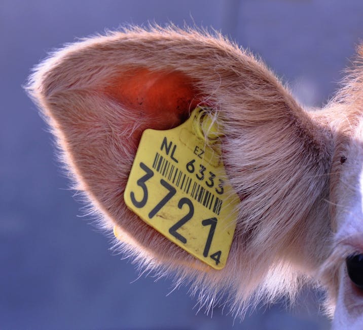
- Thoroughly clean and disinfect dehorning, castration, tattooing, and ear-tagging instruments between animals to avoid blood transfer.
-
Herd testing and management of carrier animals
- Conduct serologic or PCR testing to determine the prevalence of anaplasmosis in your herd.
- Segregate or manage positive and negative groups separately when feasible.
- In some control programs, carrier-positive cattle receive long-term chlortetracycline in feed at defined doses (e.g., around 2 mg CTC per lb body weight per day for 60 days) under veterinary supervision, aiming to reduce or clear carrier status.
-
Strategic in-feed medication
During high-risk vector seasons or when introducing susceptible animals into an endemic herd, feeding chlortetracycline at approved preventive levels (often 0.5–2 mg per lb body weight per day for cattle over 700 lb, depending on label and veterinary direction) can help suppress active infection and prevent clinical disease. This approach does not always prevent infection but can reduce deaths and severe illness. Proper intake monitoring is crucial, because underdosing reduces effectiveness.
-
Vaccination
- In some regions, killed or modified-live vaccines against Anaplasma marginale or the related species A. centrale are available. These vaccines generally do not prevent infection but can reduce the severity of clinical disease.
- Use of live A. centrale vaccine carries risks, including occasional vaccine failures and adverse effects, and must be carefully evaluated with a veterinarian.
- Some vaccines should be used with caution in late gestation because of the risk of neonatal isoerythrolysis in calves born to vaccinated cows.
-
Biosecurity and animal movement
- Avoid introducing cattle of unknown anaplasmosis status into a naive herd. New additions should ideally be tested and quarantined.
- Understand whether your region is considered endemic or free of bovine anaplasmosis and design your strategy accordingly. In disease-free areas, the priority is to prevent introduction; in endemic areas, the goal is to manage exposure, maintain herd immunity at safe levels, and reduce clinical outbreaks.
A veterinarian familiar with your local disease situation can help design a tailored prevention and control plan for Anaplasmosis in Cattle that fits your operation’s size, management style, and risk tolerance.
Frequently Asked Questions About Anaplasmosis in Cattle
Q: What exactly is Anaplasmosis in Cattle?
A: Anaplasmosis in Cattle is an infectious blood disease caused mainly by the parasite Anaplasma marginale, which infects and destroys red blood cells. This leads to severe anemia, weakness, jaundice, and sometimes death, especially in adult cattle. The disease is spread by ticks, biting flies, contaminated needles, and sometimes from cow to calf before birth.
Q: What are the main symptoms of Anaplasmosis in Cattle?
A: The key symptoms of Anaplasmosis in Cattle include fever, loss of appetite, weight loss, pale or yellow (jaundiced) mucous membranes, weakness, rapid breathing and heart rate, and sudden death after exertion. Pregnant cows may abort. Unlike some other blood diseases, the urine is usually not red or brown.
Q: How do you treat Anaplasmosis in Cattle?
A: Treatment usually involves injectable tetracycline antibiotics such as oxytetracycline under veterinary guidance. Supportive care includes minimizing stress, providing good nutrition, and, in severe cases, considering blood transfusion. Early treatment improves survival. Surviving animals often remain carriers, and in some programs, long-term tetracycline regimens are used to reduce carrier status.
Q: Can Anaplasmosis in Cattle be prevented?
A: Yes, the risk can be greatly reduced but not always completely eliminated. Key measures include tick and fly control, using a new needle for each animal, disinfecting instruments between uses, testing herds to identify carriers, strategic feeding of chlortetracycline during high-risk periods, cautious use of vaccines where available, and strict biosecurity when introducing new animals.
Q: Is Anaplasmosis in Cattle contagious from cow to cow without vectors?
A: Anaplasmosis in Cattle is considered infectious and transmissible but not directly contagious in the sense of spread by casual contact or respiratory routes. The organism must be transferred in blood, usually by ticks, biting flies, contaminated needles, or from dam to fetus. Simply grazing together does not spread the disease unless blood-sucking vectors or blood-contaminated equipment are involved.
Q: Do recovered cattle stay immune for life?
A: Cattle that survive clinical anaplasmosis usually develop long-lasting immunity to severe disease but remain carrier animals with low-level infection. They may not show signs again but can serve as sources of infection for susceptible cattle through vectors or blood transfer.
Q: Are certain cattle breeds more resistant to Anaplasmosis?
A: Evidence suggests that Bos indicus breeds (such as many zebu and tropical breeds) tend to be more resistant to Anaplasma marginale infection than Bos taurus breeds, although individuals in both types can vary. Disease severity is also affected by the strain of Anaplasma, age, and immune status.
When to Call a Veterinarian
Prompt veterinary involvement is critical when dealing with Anaplasmosis in Cattle. You should contact a veterinarian immediately if:
- Adult cattle show sudden onset of fever, weakness, pale or yellow mucous membranes, and reluctance to move, especially in a tick- or fly-infested area. These signs are classic for bovine anaplasmosis and can progress rapidly to death.
- Multiple animals in a group become ill with similar signs, or sudden deaths occur in late summer or fall. Vector-borne outbreaks often follow this pattern.
- Pregnant cows begin aborting or showing anemia and jaundice. Reproductive losses can be a key indicator of underlying anaplasmosis in naive herds.
A veterinarian will perform a physical examination, collect blood samples for smears, PCV, and laboratory tests such as ELISA or PCR, and confirm whether Anaplasmosis in Cattle is present. They can then prescribe appropriate antibiotics, advise on supportive care, and help design a herd-level control plan including testing, separation of carriers, vector control, and any permitted preventive medications or vaccines.
Because legal drug uses and withdrawal times differ between countries and markets, veterinary oversight ensures that treatment is both medically appropriate and compliant with meat and milk safety regulations. Veterinarians also help differentiate anaplasmosis from other diseases with similar signs, such as babesiosis, leptospirosis, and other causes of anemia and jaundice.
Conclusion
Anaplasmosis in Cattle is a major tick-borne blood disease that threatens cattle health and herd profitability worldwide. Caused primarily by Anaplasma marginale, it destroys red blood cells, leading to anemia, jaundice, weakness, abortions, and often death in untreated adult cattle. Its impact is amplified by the fact that recovered animals commonly become lifelong carriers, silently maintaining the parasite within the herd.
Effective management begins with recognizing the symptoms of Anaplasmosis in Cattle, such as fever, pale or yellow mucous membranes, rapid breathing, and sudden death after exertion, and then confirming the diagnosis with appropriate laboratory tests including blood smears, serology, and PCR. Understanding how to treat Anaplasmosis in Cattle with timely tetracycline therapy and supportive care can save valuable animals and reduce losses.
Long-term success depends on a well-planned prevention of Anaplasmosis in Cattle strategy: controlling ticks and biting flies, practicing strict needle and instrument hygiene, testing herds for carriers, using medicated feeds strategically, and considering vaccination where appropriate and safe. Working closely with a veterinarian to tailor these tools to your specific region and management system is the most reliable way to protect your herd from this important disease.